
articles
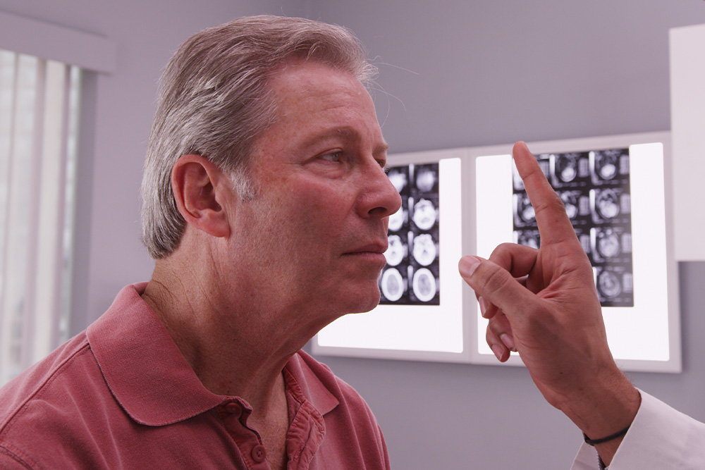
Neuro-Optometric Rehabilitation
When most people think about vision problems, they picture issues with the eyes themselves. However, many visual difficulties actually begin in the brain. After a concussion, stroke, or other brain injury, the connection between the eyes and brain can be disrupted - causing symptoms that affect balance, reading, focus, and even daily comfort.
Brain Injuries and Vision
Your visual system is complex - about 70% of the brain is involved in vision processing. After a traumatic brain injury (TBI), concussion, or neurological event such as a stroke, the pathways that coordinate eye movement, focus, and perception can become impaired.
This can result in symptoms such as:
- Blurred or double vision
- Eye strain and headaches
- Difficulty reading or focusing
- Dizziness or balance problems
- Light sensitivity
- Problems with depth perception
These visual issues often persist long after other symptoms of a brain injury have improved, making specialized vision care essential for recovery.
What Is Neuro-Optometric Rehabilitation?
Neuro-optometric rehabilitation is a specialized form of vision therapy designed to retrain how the brain and eyes work together. Unlike traditional eye care that focuses on eyesight alone, this therapy targets the neurological processes behind vision. Our doctor evaluates how the eyes and brain communicate and creates a personalized treatment plan to rebuild these visual pathways. The goal is to restore comfort, coordination, and clarity - improving overall visual function and quality of life.

Our eyes are extremely delicate, yet they can be subjected to harsh conditions and other environmental factors that affect their health. One of the problems that can affect our eyes is an accumulation of dirt, debris and bacteria on the eyelids. This can cause a range of issues, including stopping tear film from reaching the eyes and being properly dispersed over their surface – which is necessary to keep them healthy and comfortable. Fortunately, a new solution called Blephex® can help.
What is Blephex®?
Blephex® is a handheld electro-mechanical device that is applied to the margins of the eyelids with the purpose of cleaning them and improving the effectiveness with which tear film flows onto the surface of the eyes.
Blephex® has a disposable, surgical-grade sponge tip which rapidly oscillates to create a cleaning action. Before the sponge tip is placed onto the eyes, it is soaked in a gentle exfoliating solution. This solution provides soft abrasion to help remove dead skin cells and debris that could be irritating the eyes and interrupting tear film progression. The Blephex® device is manually applied to the eyes and moved gently across the eyelids, with the entire, painless process taking approximately 6 to 8 minutes per eye. A different sponge is used on each eye, ensuring that no bacteria is passed between them. After the procedure, patients are given instructions on how to maintain the cleanliness of their eyelids with daily/nightly eyelid hygiene at home.
Most patients experience a significant improvement in tear film production and dispersal, and a reduction in unpleasant symptoms that they may have been experiencing within 48 hours of their treatment. While a single treatment is normally enough to produce excellent results, many patients are advised to have Blephex® every 4-6 months.

When it comes to maintaining your eye health, regular check-ups and screenings play a crucial role. One such screening method that has revolutionized the field of optometry is retinal imaging testing. This non-invasive procedure allows optometrists to capture detailed images of the retina, providing valuable insights into the overall health of your eyes.
The Importance of Retinal Imaging Testing
By examining the retina, which is the thin layer of tissue at the back of your eye responsible for capturing light and transmitting visual signals to the brain, optometrists can gain valuable insights into your eye health. Retinal imaging testing allows for the early detection of various eye conditions even before noticeable symptoms occur. This early detection is crucial as it enables prompt treatment and intervention, potentially preventing irreversible vision loss.
Advanced Retinal Imaging Technology
Retinal imaging testing has been made possible by the advancements in technology, specifically Optical Coherence Tomography (OCT) and Optos imaging. OCT is a non-invasive imaging technique that uses light waves to capture high-resolution cross-sectional images of the retina. It provides detailed information about the layers of the retina, helping eye doctors identify and monitor various eye conditions.
Optos imaging utilizes ultra-widefield retinal technology to capture a panoramic image of the retina. This technology allows for a more comprehensive view of the retina, including the periphery. Dilating drops are not necessary with Optos, making the process more convenient for patients.

Dry eye is a common ocular condition that occurs when the eyes do not produce enough tears or when the tears evaporate too quickly. This can lead to discomfort, redness, blurred vision, and even damage to the surface of the eyes. Understanding the causes and symptoms of dry eye is crucial provide early detection and effective treatment.
The Importance of Dry Eye Advanced Diagnostic Testing
Early detection of dry eye is crucial for preventing further progression of the condition and improving patient outcomes. Without proper diagnosis and treatment, dry eye can cause significant discomfort and affect daily activities. Additionally, chronic dry eye can lead to corneal ulcers, infections, and even vision loss. By accurately identifying and addressing dry eye in its early stages, optometrists can provide timely interventions and prevent complications.
TearLab
One of the advanced diagnostic tools available for dry eye is TearLab. TearLab is a non-invasive test that measures the osmolarity of tears, which is an indicator of tear film stability. This test provides valuable information about the quality and quantity of tears, allowing healthcare professionals to accurately diagnose and monitor dry eye. By analyzing the osmolarity of tears, TearLab helps identify the severity of dry eye and guides treatment decisions. It is a quick and painless procedure that can be performed in a clinical setting.
InflammaDry
Inflammation plays a significant role in dry eye, and identifying the presence of inflammation is crucial for effective treatment. InflammaDry is a diagnostic tool that detects elevated levels of matrix metalloproteinase 9 (MMP-9), an inflammatory marker, in tears. By measuring MMP-9, InflammaDry helps optometrists differentiate between inflammatory and non-inflammatory dry eye. This information is essential for tailoring treatment plans and determining the most appropriate therapies for each patient.

Corneal refractive therapy, also known as CRT, is a simple, painless treatment for refractive eye errors like myopia and has two core benefits. First, it can be used to help patients see clearly during the day without using glasses or contact lenses, giving them the freedom and flexibility that they need to live life to the fullest. Second, CRT has been shown to help slow the progression of myopia, keeping prescriptions under control and potentially reducing the likelihood of patients developing serious eye health problems associated with high myopia in the future.
Here’s everything that you need to know about corneal refractive therapy and what it means for you.
Understanding refractive eye problems
Refractive eye problems like nearsightedness, farsightedness and astigmatism are extremely common, with nearsightedness – also known as myopia – being the most common of all. Patients with myopia can see nearby objects clearly, but those further away become progressively more blurred. Refractive eye errors occur when the shape of the clear dome covering the front part of the eye, called the cornea, impair the light-bending and focusing process in your eyes. This leads to the light ending up in the wrong place inside the eye, and the message that is sent to our brain from our eyes is muddled, causing blurred vision.
What is corneal refractive therapy?
Corneal refractive therapy was initially developed as a treatment to correct and slow the progression of nearsightedness. However, it has also been found to be effective at controlling other refractive errors, including farsightedness, astigmatism and an age-related refractive condition called presbyopia.
CRT is a non-invasive, painless and straightforward method of correcting patient vision so that they don’t need to wear contacts or glasses, and they don’t need laser vision correction surgery to see clearly. CRT uses special contact lenses that are worn overnight and apply light pressure to the cornea in order to reshape it so that light is refracted correctly, and the image sent from the eyes to the brain is clear. The cornea is able to retain this new shape even after the contact lenses are removed the next morning, meaning that you can continue to see clearly for several hours. The more consistently you wear your CRT lenses overnight, the longer your eyes will learn to retain their new shape and eventually, patients can enjoy up to 48 hours of clear vision without using prescription lenses. However, the effects aren’t permanent so if you stop wearing the lenses, your vision will gradually return back to normal over the course of a few days.
Slowing the progression of myopia with corneal refractive therapy
Another key benefit of CRT is that it can actually help to slow the progression of myopia. Most people who are nearsighted find that their eyesight gets progressively worse as they get older. This deterioration may not be rapid, but it can end in patients requiring high prescriptions. Studies have found that patients who have high myopia are more likely to develop serious eye problems in the future, including glaucoma, macular degeneration, cataracts and a detached retina. Regular use of your corneal refractive therapy lenses could help keep your prescription stable and lower your risk of developing these problems.

Newton™
Newton™, formerly Neurolens®, is the first and only prescription lenses that include an element of contoured prism in their design. This prism is designed to bring the patient’s eyes into more equal alignment, and this should help to provide relief from the symptoms that are associated with several eye misalignment conditions, including digital eye strain and binocular vision dysfunction.
What is digital eye strain?
Digital eye strain is the name given to describe a group of symptoms that can occur when someone spends long periods of time using digital devices. Since using digital devices requires the eyes to work harder than normal and we don’t always position our devices the perfect distance away, it can lead to issues such as eye pain, dry and irritated eyes, eye fatigue, light sensitivity, and blurred vision. Unsurprisingly, the number of people who are experiencing digital eye strain has grown significantly over the last few years and is expected to continue to do so.
What is binocular vision dysfunction?
Binocular vision dysfunction, also known as BVD for short, is another eye condition but is one that is very misunderstood. Binocular vision dysfunction occurs when the eyes aren’t perfectly aligned, causing your brain and eyes to work harder than normal in order to create a clear visual image and remain focused. This places pressure on the trigeminal nerve, which is the nerve that is responsible for the majority of the sensations that we experience in our head and back. BVD can often manifest as other things owing to the huge range of symptoms that are associated with the condition. These can include, but aren’t limited to:
Blurred vision
Headaches/migraines
Double vision
Motion sickness
Vertigo
Dizziness
Anxiety
Many people don’t think to visit an eye doctor when they are experiencing these symptoms, but all can occur simply because the eyes are out of alignment.

Medical eye exams are comprehensive evaluations of the health and function of the eyes, essential for maintaining optimal vision and preventing potential eye conditions. Regular medical eye exams are fundamental for proactive eye health management and ensuring ongoing visual wellness.
What is a Medical Eye Exam?
A medical eye exam is a comprehensive examination of your eyes conducted by an optometrist. This examination goes beyond a routine vision test and delves into the overall health of your eyes. During a medical eye exam, various tests will be performed to assess your visual acuity, peripheral vision, eye movement, and the health of your eye structures.
Medical eye exams can detect early signs of eye conditions that may not present noticeable symptoms in their initial stages. By diagnosing these conditions early, you have a greater chance of successful treatment and preventing further deterioration of your vision.
Medical eye exams can also identify underlying health conditions that may manifest in your eyes, providing an opportunity for early intervention and management.
Common Conditions Detected During a Medical Eye Exam
During a medical eye exam, several common eye conditions can be detected, even before noticeable symptoms occur. One such condition is age-related macular degeneration (AMD), which affects the central part of the retina responsible for sharp, central vision. By examining the retina, your optometrist can identify early signs of AMD and recommend appropriate treatment to slow its progression.
Another condition that can be detected during a medical eye exam is cataracts. Cataracts occur when the lens of your eye becomes cloudy, leading to blurred vision. Through a comprehensive examination, your eye doctor can assess the severity of your cataracts and recommend the most suitable treatment option, which may include surgery to remove the cloudy lens.
Hypertension is a common health condition that can have serious consequences if left untreated. The health of your eyes can provide valuable insights into your cardiovascular health. During a medical eye exam, an optometrist can observe changes in the blood vessels of your retina, such as narrowing or leaking. These changes can indicate underlying hypertension.

Astigmatism is a common refractive error that occurs when the cornea or lens of the eye is not perfectly spherical. Instead of having a uniform, rounded shape, the cornea or lens has a slightly more curved or irregular shape, resembling more of an oval or football-like appearance.
This irregular shape causes light to bend or refract unevenly as it enters the eye, leading to blurred or distorted vision. Astigmatism is often present from birth, but it can also develop later in life due to various factors, such as eye injuries, certain medical conditions, or the natural aging process.
Common Symptoms of Astigmatism
If you have astigmatism, you may experience a range of symptoms that can impact your daily life. Some of the most common symptoms of astigmatism include:
Blurred or distorted vision, especially at a distance or when reading
Difficulty seeing clearly at night or in low-light conditions
Frequent headaches or eye strain
Sensitivity to glare or bright lights
Double vision or the appearance of halos around lights
These symptoms can vary in severity and may become more pronounced over time, especially if the astigmatism is left uncorrected.

Presbyopia is a natural and inevitable part of aging that affects the eyes' ability to focus on close objects. Unlike other vision conditions, presbyopia isn’t caused by the shape of the eye or structural abnormalities but is instead a result of the eye’s lens losing its flexibility over time.
What is Presbyopia?
Presbyopia is a refractive error, meaning it affects how the eyes bend (or refract) light to focus it on the retina. Unlike nearsightedness (myopia) or farsightedness (hyperopia), which are caused by the shape of the eyeball or cornea, presbyopia is due to the aging of the eye's lens. Over time, the lens becomes less flexible, making it difficult to focus on nearby objects. Presbyopia isn’t a disease or an abnormality—it’s simply a natural consequence of the aging process.
What Causes Presbyopia?
The lens inside the eye changes shape to focus light on the retina, allowing us to see clearly at varying distances. When viewing nearby objects, the lens thickens and curves to increase its refractive power.
As we age, however:
Lens Stiffening: The lens loses elasticity, making it harder to bend and adjust for near vision.
Weakened Ciliary Muscles: The muscles that help change the shape of the lens may lose their strength over time, further reducing the eye’s ability to focus on close objects.
Structural Changes: The lens also grows thicker and less transparent with age, which contributes to reduced focusing ability.
These changes are gradual, typically starting in a person’s 30s and becoming noticeable by their early to mid-40s.

Dry eyes are one of the most common conditions that can affect our eyes and is estimated to affect millions of Americans. As you’ve probably guessed, dry eyes occur when tears fail to provide enough natural lubrication for the eyes to be comfortable and healthy. Exactly what causes dry eyes can vary significantly, from side effects from medications to prolonged computer use. What is clear is that while the condition isn’t sight-threatening, it can make day to day life much harder than it needs to be. Fortunately, there are treatments that can help, and arguably one of the most effective is Lipiflow.
What is Lipiflow?
Lipiflow is a new technological solution that addresses the underlying cause of your dry eyes, rather than simply treating the symptoms. It is most effective at helping patients whose dry eyes are caused by meibomian gland dysfunction – a condition characterized by problems with the way that the meibomian glands produce the oil that forms an essential part of our tear film. The meibomian glands can become less productive, or in some cases, even blocked by hardened oil deposits. This prevents the oil from reaching your tear film, making it less effective. Lipiflow targets the meibomian glands, warming them to break down oily blockages and massaging your eyes to make sure that the oil, and then the tear film, is evenly dispersed. This helps to combat the symptoms associated with dry eyes, which can include:
Eye fatigue
Dry, scratchy and uncomfortable eyes
Blurred vision
Sensitivity to light
Difficulty wearing contact lenses
Your eye doctor will be able to advise you if Lipiflow has the potential to be a suitable solution for your dry eyes.

© 2026 Linden Optometry, A P.C.. All rights Reserved - Accessibility Statement - Privacy Policy - Sitemap
Powered by